Isopod iridescent virus 31
IIV-31
Table of Contents
Signs and Symptoms
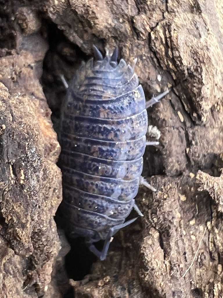
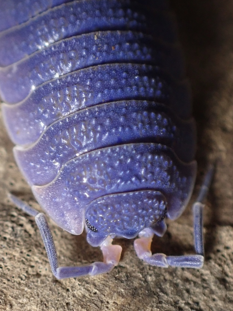
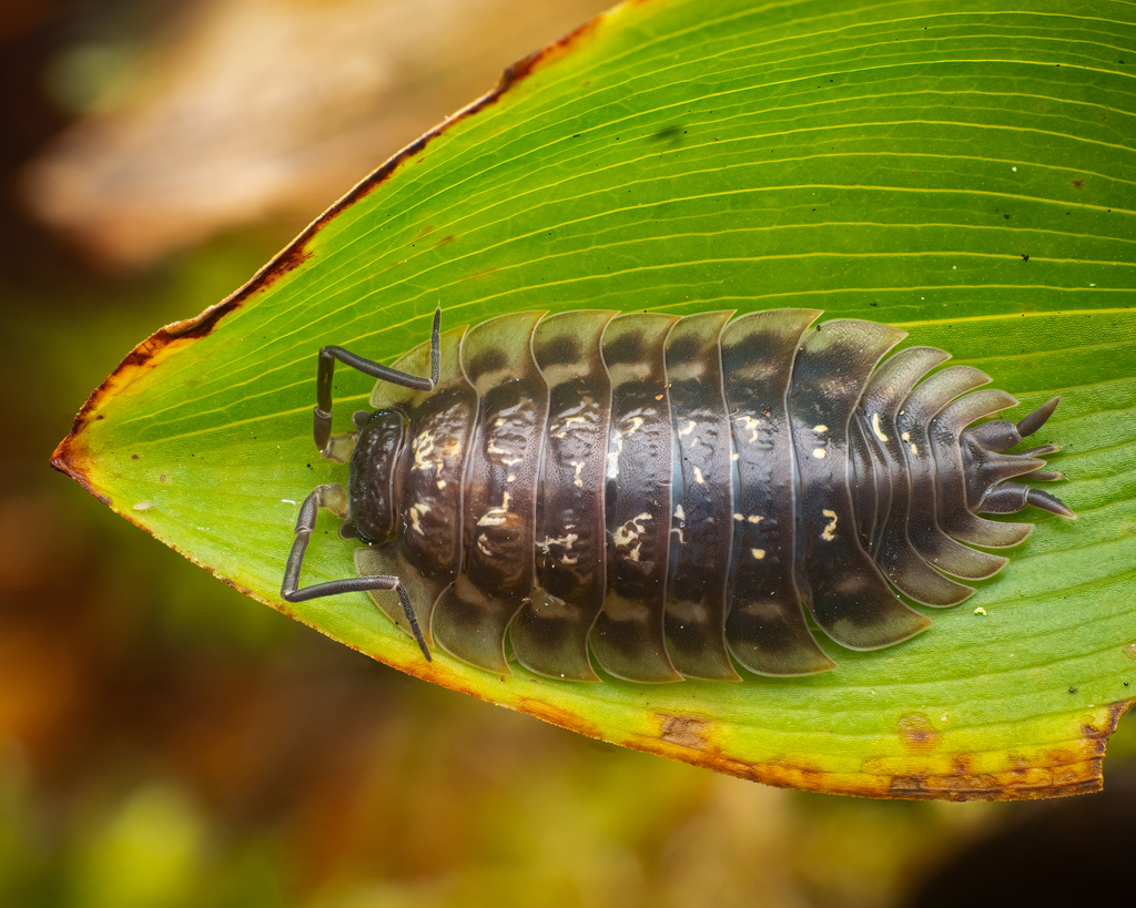
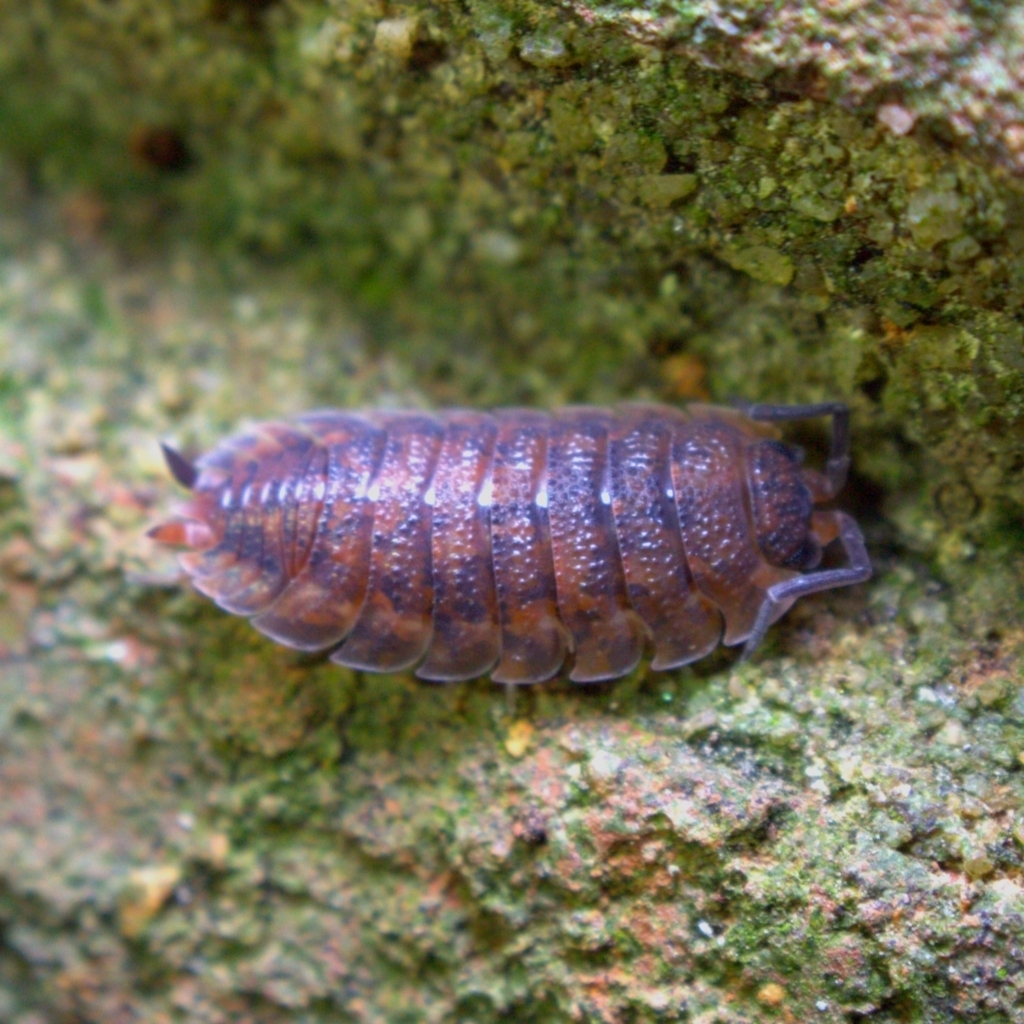
The infection is not immediately visible, but turns the isopod blue or purple over time. In earlier stages, the infected individual can look just slightly purple or blue. 4 1
Progression
The blue colour is first visible in the non-pigmented parts of the isopod. This is often the ventral surface (tummy) but it depends on the species. 4
Later on, the more pigmented parts (such as the back) become blue or purple and often times purple spots form on the dorsal surface (back). The intensity of the colouration is very variable between individuals and also depends on the viral burden. 4
In the later stages, organs can contain the virus, too. The animals get more lethargic and less responsive to stimuli. They probably also eat less. 4
Not every animal lives until the later stages. A study found that some die after as little as two days and others live up to 60+ days. 4
When isopod were injected with the virus, they showed discolouration after 11-14 days and died 17-20 days after showing discolouration. It was suggested, that lower temperatures lead to the infected individuals living longer. 4
Reason for the colour
The colour stems from virus particles in the infected cells. The particles have a para-crystalline structure and reflect the light in a way that makes it look blue. 4
Transmission
As per os infection almost invariably requires ingestion of very large doses of particles, cannibalism or predation of infected individuals is considered a likely mechanism of transmission in […] terrestrial isopods. 1
It is suggested that cannibalism massively contributes to the spread of the disease. 1
Though the same paper suggests that IIVs in general are not necessarily transmitted through ingestion only (healthy individuals getting infected through eating dead infected ones) but also other mechanisms such as wounding. 1
Intra- and interspecific aggression between individuals resulting in wounding has been identified as an important factor influencing IIV transmission in natural populations of isopods and laboratory populations of mosquitoes. 1
This is especially important for rather aggressive species such as some Porcellios.
Doubling the density of Porcellio scaber populations resulted in an insignificant change in the prevalence of infection by IIV-31, whereas introducing an equal density of Porcellio laevis was accompanied by a threefold increase in infection. 1
Under laboratory ambient, the virus remained infective for five days after the death of the host isopod. Other IIVs also showed reduced activity in very dry soil, which suggests that a dry habitat could help slow down infection. 1
Exposure to solar UV light resulted in a very rapid inactivation of IIV-6 in water 1
UV light was only tested on VII-6 but it is quite possible this method could be used on IIV-31, too.
Contributing factors
As established above, aggression can contribute to a faster spreading.
There was also a seasonality of infection in wild terrestrial isopod infections observed. During dry months, the isopods tend to travel smaller distances and stay aggregated in big numbers. This prevents the spread over large distances that are more likely to occur in wet months. 1
It is also possible that (as stated above) the dry environment leads to a faster inactivation of the virus in the soil.
Prevalence
Many different species and genera of isopods are known to be infected by this virus. There also is a parasitic nematode that can carry this virus and is known to parasite on isopods.1 2
Infections are usually contained to relatively small sites of a few m2. The count of infected individuals per site is difficult to estimate, because the colour only starts showing later on. 4
Species that are known to be hosts
| Mermithidae sp. 2 (Nematode) |
| Alloniscus balssi 7Alloniscus balssi |
| Androniscus dentiger 4 |
| Armadillidium decorum 6 |
| Armadillidium vulgare 3 |
| Burmoniscus kathmandia 7 |
| Haplophthalmus danicus 4 |
| Haplophthalmus mengii 4 |
| Hyloniscus riparius 4 |
| Ligidium hypnorum 4 |
| Ligidium koreanum 7 |
| Littorophiloscisa nipponensis 7 |
| Oniscus asellus 4 |
| Philoscia affinis 6 |
| Philoscia muscorum 6 |
| Porcellio dilatatus 4 |
| Porcellio laevis 1 |
| Porcellio scaber 3 |
| Porcellio siculoccidentalis 6 |
| Porcellio spinicornis 4 |
| Trichoniscoides albidus 4 |
| Trichoniscoides helveticus 4 |
| Trichoniscus panormidensis 6 |
| Trichoniscus pusillus 4 |
| Venezillo obscurus 7 |
Pictures